Home

Plânge nichel Analitic cell division microscope microscope obosit Medalie Nevinovat

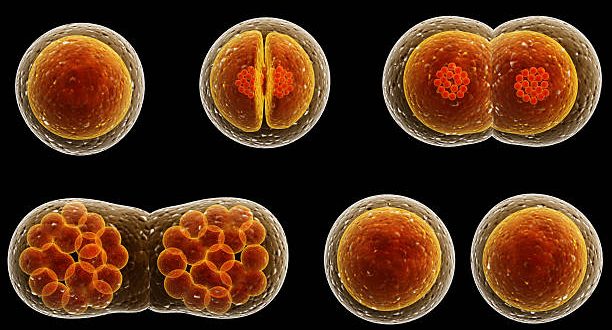
Biology concept. Cell division under the microscope. 3d illustration Stock  Photo by ©urfingus 304694846
Biology concept. Cell division under the microscope. 3d illustration Stock Photo by ©urfingus 304694846

Microscopic images of chromosomes at different stages of cell division... |  Download Scientific Diagram
Microscopic images of chromosomes at different stages of cell division... | Download Scientific Diagram

Cell Division and Cell Cycle under the microscope. Stock Photo | Adobe Stock

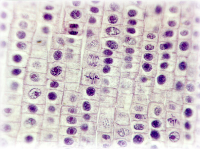
Cell Microscopy Lesson Objectives (L.O.) To calculate the mitotic index  within a field of view 31 st January 2006 To be able to measure cells in  interphase. - ppt download
Cell Microscopy Lesson Objectives (L.O.) To calculate the mitotic index within a field of view 31 st January 2006 To be able to measure cells in interphase. - ppt download

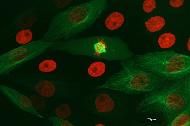
Science Source Images - Cell Division - Immunofluorescent Microscopy  Immunofluorescent light micrograph of a cell (center) during the metaphase  stage of mitosis (cell division). It is surrounded by interphase (resting)  cells. During
Science Source Images - Cell Division - Immunofluorescent Microscopy Immunofluorescent light micrograph of a cell (center) during the metaphase stage of mitosis (cell division). It is surrounded by interphase (resting) cells. During

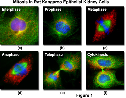
Molecular Expressions Cell Biology: Mitosis with Fluorescence Microscopy

How to See the Cell Cycle Through Your Microscope

Human cells: Cell cycle temperature control | Cherry Biotech

How to Identify Stages of Mitosis Within a Cell Under a Microscope |  Sciencing
How to Identify Stages of Mitosis Within a Cell Under a Microscope | Sciencing

Cell Division: Types, Stages & Processes : Plantlet

Biology Concept Cell Division Under The Microscope Isolated On A White 3d  Illustration Stock Photo - Download Image Now - iStock
Biology Concept Cell Division Under The Microscope Isolated On A White 3d Illustration Stock Photo - Download Image Now - iStock
Mic-UK: Mitosis - cell division

Cell Division Cell Cycle Plant Under Stock Photo 484811614 | Shutterstock

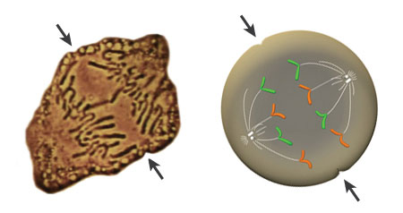
Cell division observed through the microscope (left) is redrawn to show the  action of chromosomes (right). Arrows indicate the axis along which the cell  divides. | Learn Science at Scitable
Cell division observed through the microscope (left) is redrawn to show the action of chromosomes (right). Arrows indicate the axis along which the cell divides. | Learn Science at Scitable

Mitosis Through the Microscope: Advances in Seeing Inside Live Dividing  Cells | Science
Mitosis Through the Microscope: Advances in Seeing Inside Live Dividing Cells | Science

Pin on School
Pin on School

A-D Transmission electron microscopy (TEM) micrographs of cell division...  | Download Scientific Diagram
A-D Transmission electron microscopy (TEM) micrographs of cell division... | Download Scientific Diagram

Mitosis Through the Microscope: Advances in Seeing Inside Live Dividing  Cells | Science
Mitosis Through the Microscope: Advances in Seeing Inside Live Dividing Cells | Science

Cell Division And Cell Cycle Under The Microscope. Stock Photo, Picture And  Royalty Free Image. Image 161785649.
Cell Division And Cell Cycle Under The Microscope. Stock Photo, Picture And Royalty Free Image. Image 161785649.
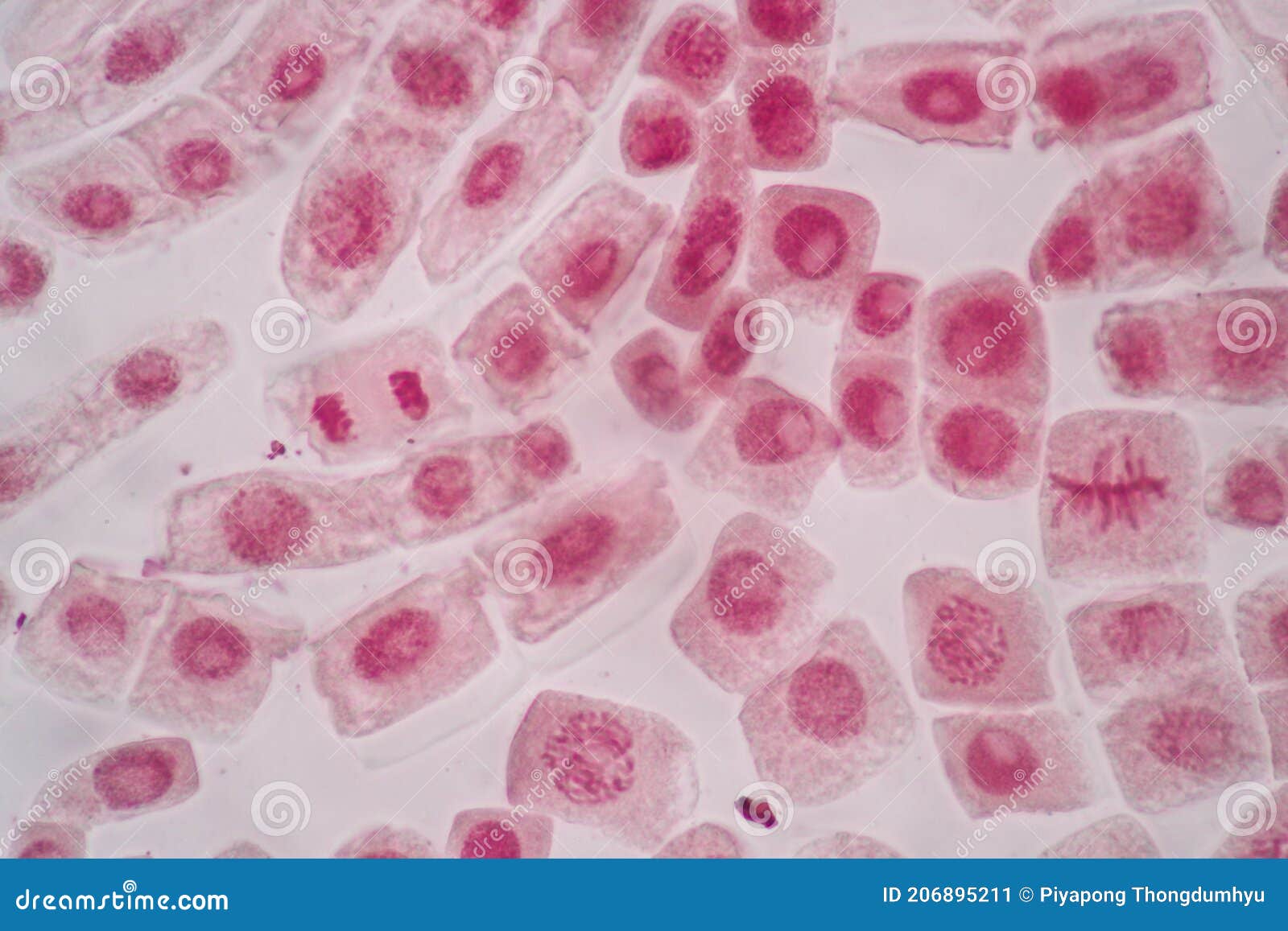
Cell Division and Cell Cycle Under the Microscope. Stock Image - Image of  centromeres, membrane: 206895211
Cell Division and Cell Cycle Under the Microscope. Stock Image - Image of centromeres, membrane: 206895211

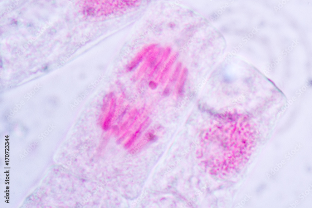
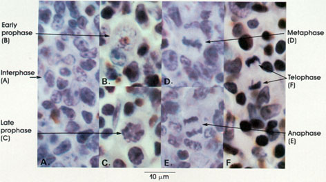
Anatomy Atlases: Atlas of Microscopic Anatomy: Section 1 - Cells

Cell Division, Scientific Medical Experiment Under Microscope, 3d Rendering  Stock Illustration - Illustration of division, nucleus: 139889697
Cell Division, Scientific Medical Experiment Under Microscope, 3d Rendering Stock Illustration - Illustration of division, nucleus: 139889697

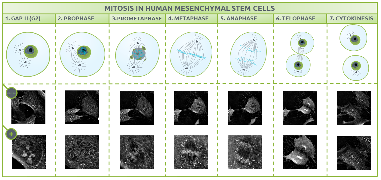
Nanolive imaging captures discrete phases of mitosis in spectacular detail

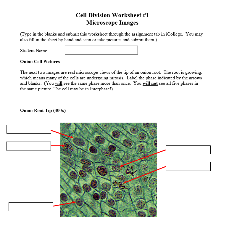
Solved Cell Division Worksheet #1 Microscope Images (Type in | Chegg.com

cell division on contrast microscope - YouTube