Home

Fulger Manual Sudan bright field live cell imaging microscope analiză completa astăzi

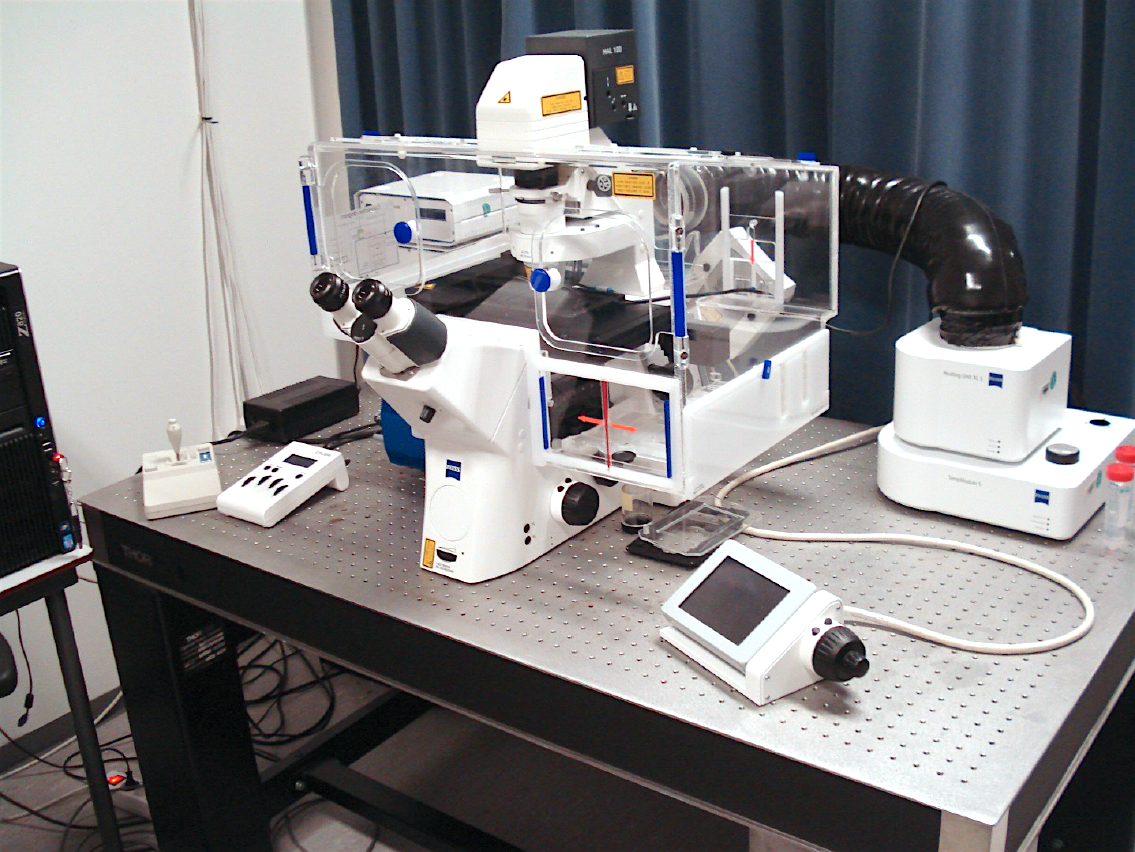
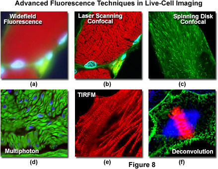
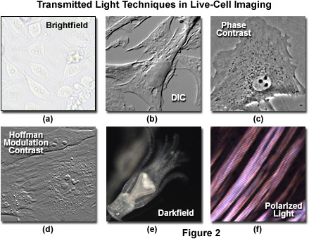
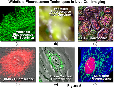
ZEISS Microscopy Online Campus | Live-Cell Imaging | Microscopy Techniques

Automated Bright Field Segmentation of Cells and Vacuoles Using Image  Processing Technique - Chiang - 2018 - Cytometry Part A - Wiley Online  Library
Automated Bright Field Segmentation of Cells and Vacuoles Using Image Processing Technique - Chiang - 2018 - Cytometry Part A - Wiley Online Library

Automated Bright Field Segmentation of Cells and Vacuoles Using Image  Processing Technique - Chiang - 2018 - Cytometry Part A - Wiley Online  Library
Automated Bright Field Segmentation of Cells and Vacuoles Using Image Processing Technique - Chiang - 2018 - Cytometry Part A - Wiley Online Library

Microscopy – Membrane Enzymology

ZEISS Microscopy Online Campus | Live-Cell Imaging | Microscopy Techniques

Live cell imaging of the HCT116 cells following treatment with 3a, 3c and  3d using bright field optical microscopy (BF) and fluorescence microscopy  (DAPI filter).
Live cell imaging of the HCT116 cells following treatment with 3a, 3c and 3d using bright field optical microscopy (BF) and fluorescence microscopy (DAPI filter).

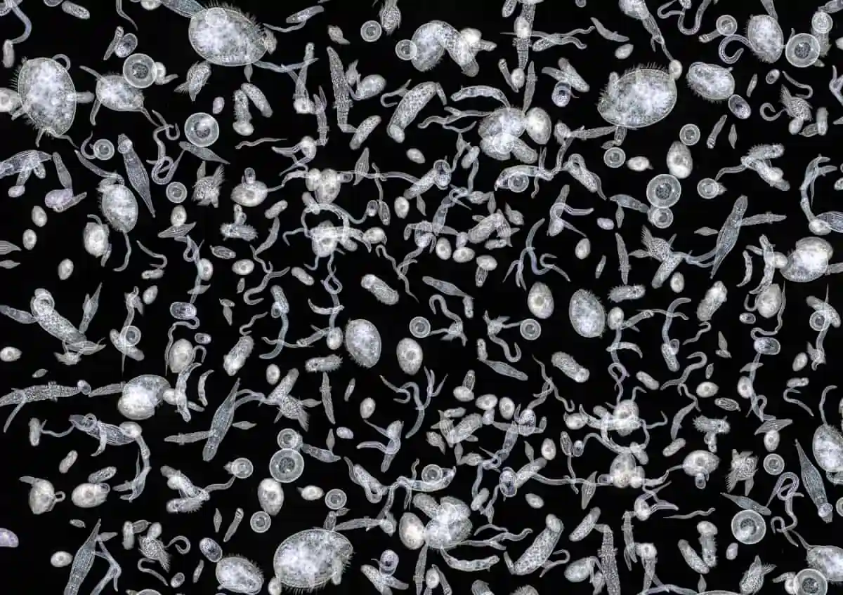
What are the differences between brightfield, darkfield and phase contrast?  | Microbehunter Microscopy
What are the differences between brightfield, darkfield and phase contrast? | Microbehunter Microscopy
What Is Live Cell Imaging?

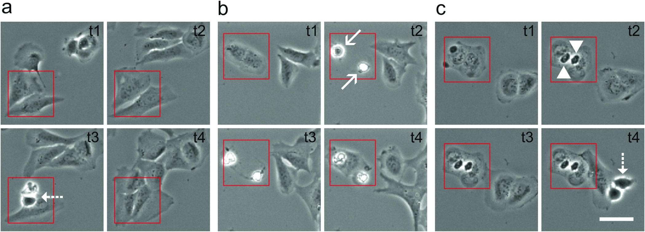
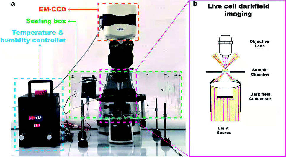
Continuous live cell imaging using dark field microscopy - Analytical  Methods (RSC Publishing) DOI:10.1039/D2AY00043A
Continuous live cell imaging using dark field microscopy - Analytical Methods (RSC Publishing) DOI:10.1039/D2AY00043A
Darkfield And Phase Contrast Microscopes

Tips for Running a Successful Live Cell Imaging Experiment | Molecular  Devices
Tips for Running a Successful Live Cell Imaging Experiment | Molecular Devices

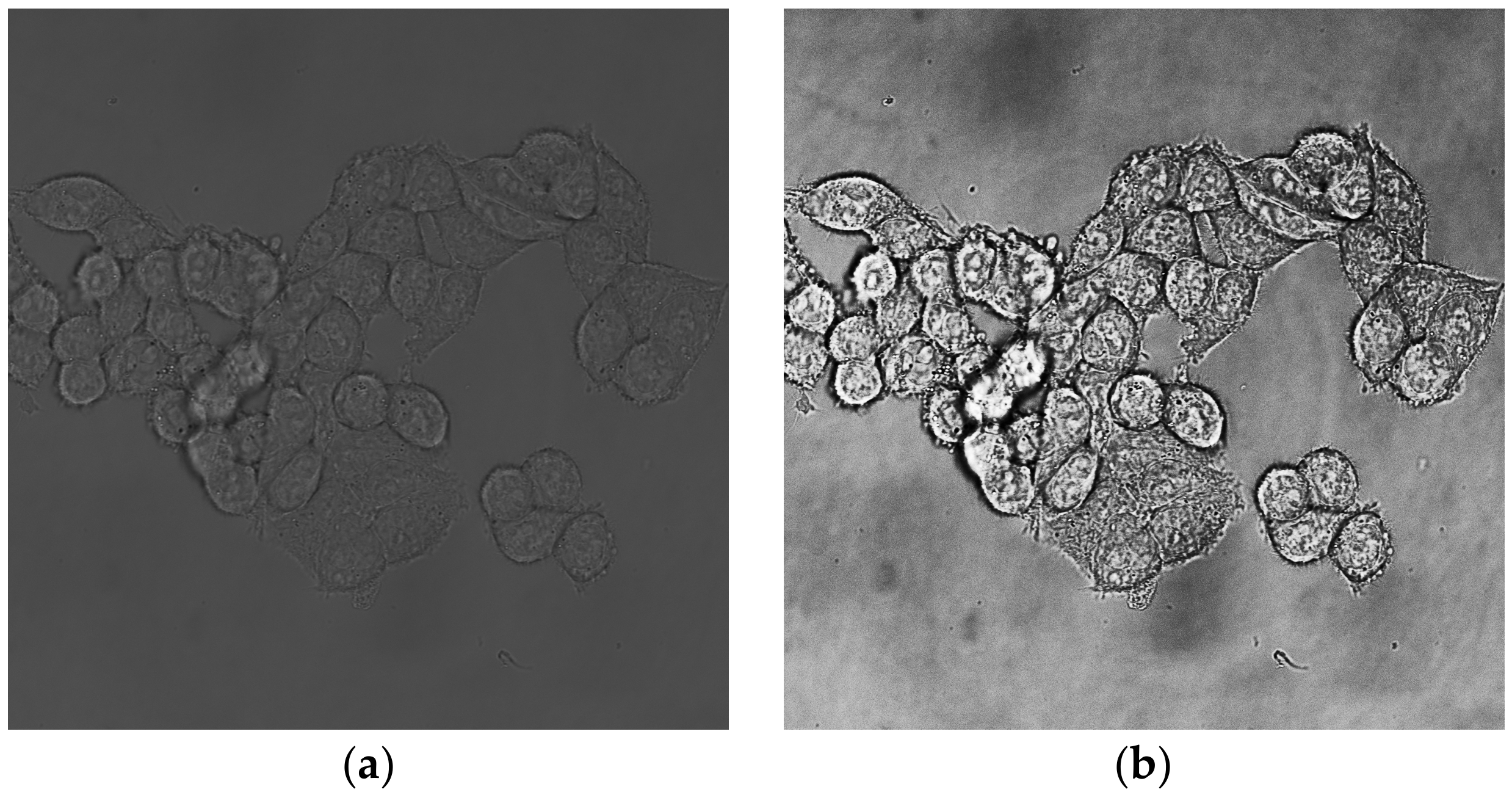
MPs | Free Full-Text | Segmentation of Total Cell Area in Brightfield  Microscopy Images
MPs | Free Full-Text | Segmentation of Total Cell Area in Brightfield Microscopy Images

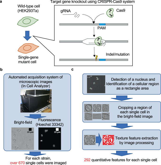
Machine learning approach for discrimination of genotypes based on bright-field  cellular images | npj Systems Biology and Applications
Machine learning approach for discrimination of genotypes based on bright-field cellular images | npj Systems Biology and Applications

A portable low-cost long-term live-cell imaging platform for biomedical  research and education - ScienceDirect
A portable low-cost long-term live-cell imaging platform for biomedical research and education - ScienceDirect

Bright field (left) and fluorescence (right) images of the HEK cells... |  Download Scientific Diagram
Bright field (left) and fluorescence (right) images of the HEK cells... | Download Scientific Diagram

Light-induced cell damage in live-cell super-resolution microscopy |  Scientific Reports
Light-induced cell damage in live-cell super-resolution microscopy | Scientific Reports

Bright Field Microscopy as an Alternative to Whole Cell Fluorescence in  Automated Analysis of Macrophage Images | PLOS ONE
Bright Field Microscopy as an Alternative to Whole Cell Fluorescence in Automated Analysis of Macrophage Images | PLOS ONE
Bright field cell imaging system - All medical device manufacturers

Counting unstained, confluent cells by modified bright-field microscopy |  BioTechniques
Counting unstained, confluent cells by modified bright-field microscopy | BioTechniques

ZEISS Microscopy Online Campus | Live-Cell Imaging | Microscopy Techniques

What Is Widefield Imaging?

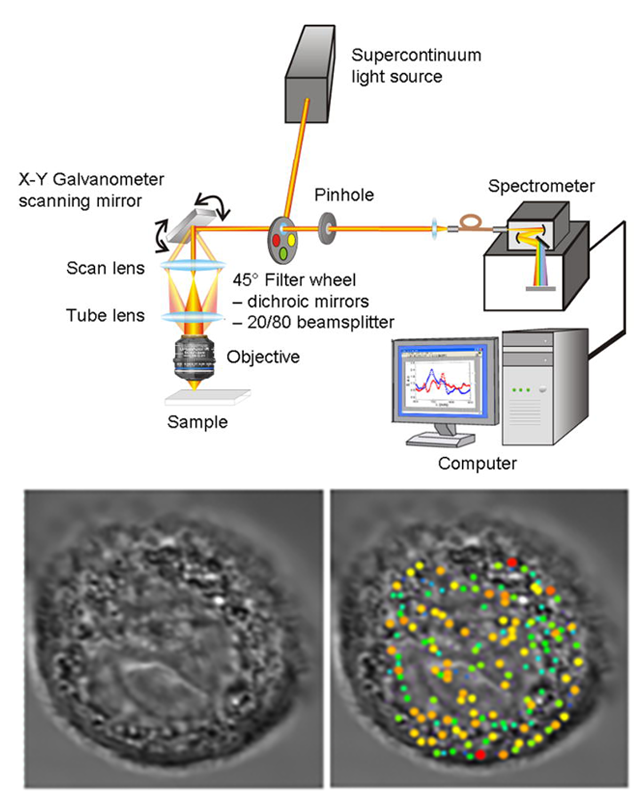
Research - Center for Advanced Biomedical Imaging and Photonics | HMS -  BIDMC
Research - Center for Advanced Biomedical Imaging and Photonics | HMS - BIDMC

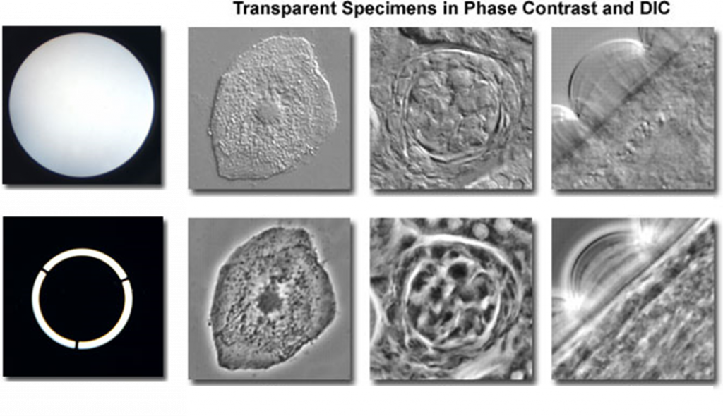
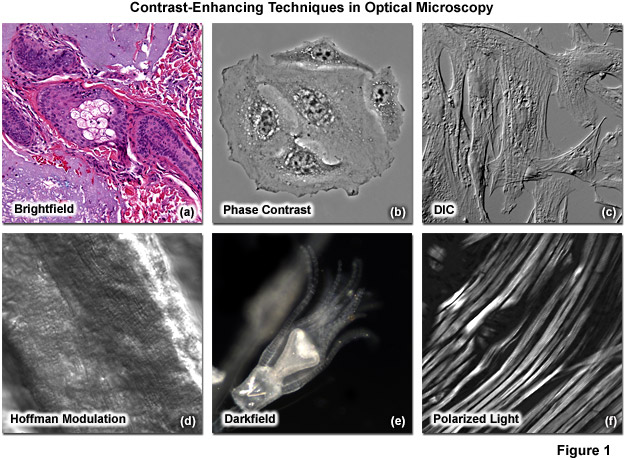
ZEISS Microscopy Online Campus | Microscopy Basics | Enhancing Contrast in  Transmitted Light
ZEISS Microscopy Online Campus | Microscopy Basics | Enhancing Contrast in Transmitted Light

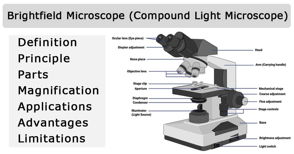
Brightfield Microscope (Compound Light Microscope)- Definition, Principle,  Parts
Brightfield Microscope (Compound Light Microscope)- Definition, Principle, Parts

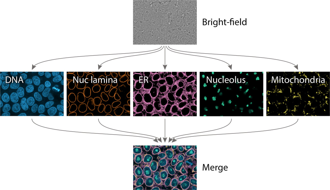
Extracting multiple cellular structures from single bright-field microscopy  images - ALLEN CELL EXPLORER
Extracting multiple cellular structures from single bright-field microscopy images - ALLEN CELL EXPLORER

Cell growth and imaging quality (a) Bright field images of one imaging... |  Download Scientific Diagram
Cell growth and imaging quality (a) Bright field images of one imaging... | Download Scientific Diagram

Live Cell Imaging
Live Cell Imaging

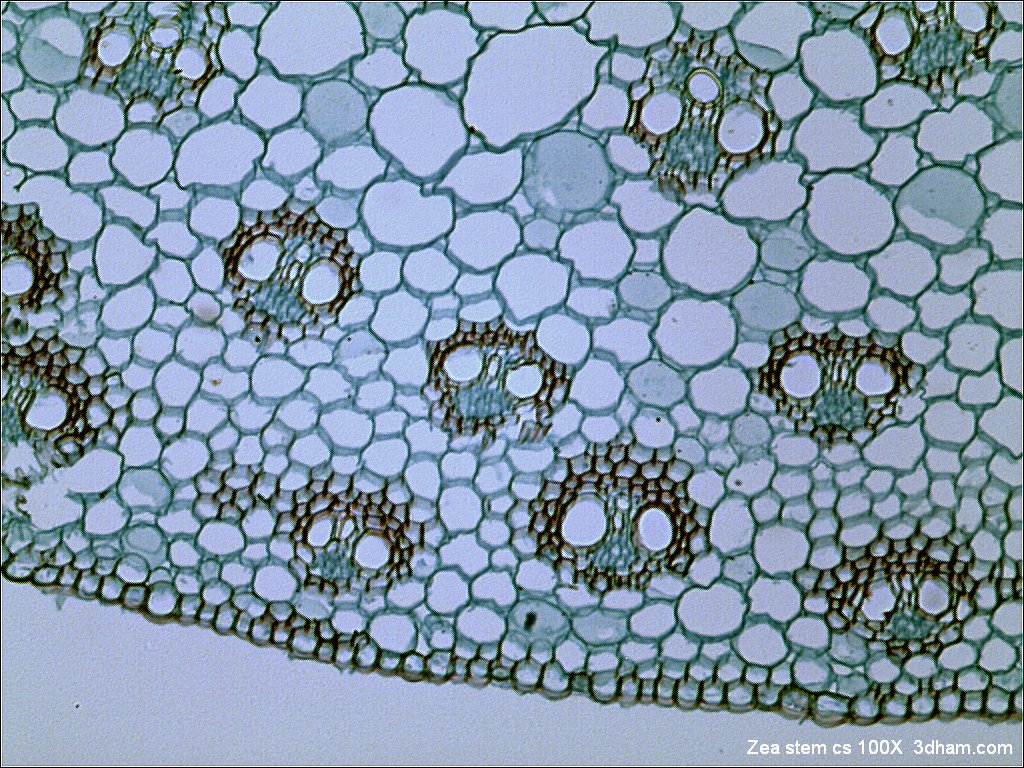
Bright-field microscopy - Wikipedia